Prefrontal neural geometry of learned cues guides motivated behaviours
TL;DR
The dorsomedial prefrontal cortex (dmPFC) encodes appetitive and aversive values of learned stimuli, with subpopulations representing valence and salience orthogonally. This neural geometry dynamically shapes motivated behaviors in mice.
Key Takeaways
- •dmPFC populations primarily encode the appetitive and aversive values of learned stimuli.
- •Subpopulations in the dmPFC represent valence and salience along orthogonal information axes.
- •The geometry of dmPFC neuronal representations dynamically guides appetitive and aversive motivated behaviors.
Tags
Abstract
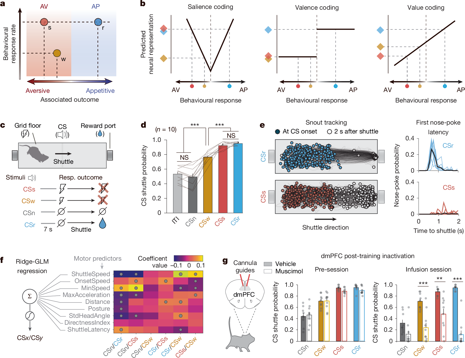
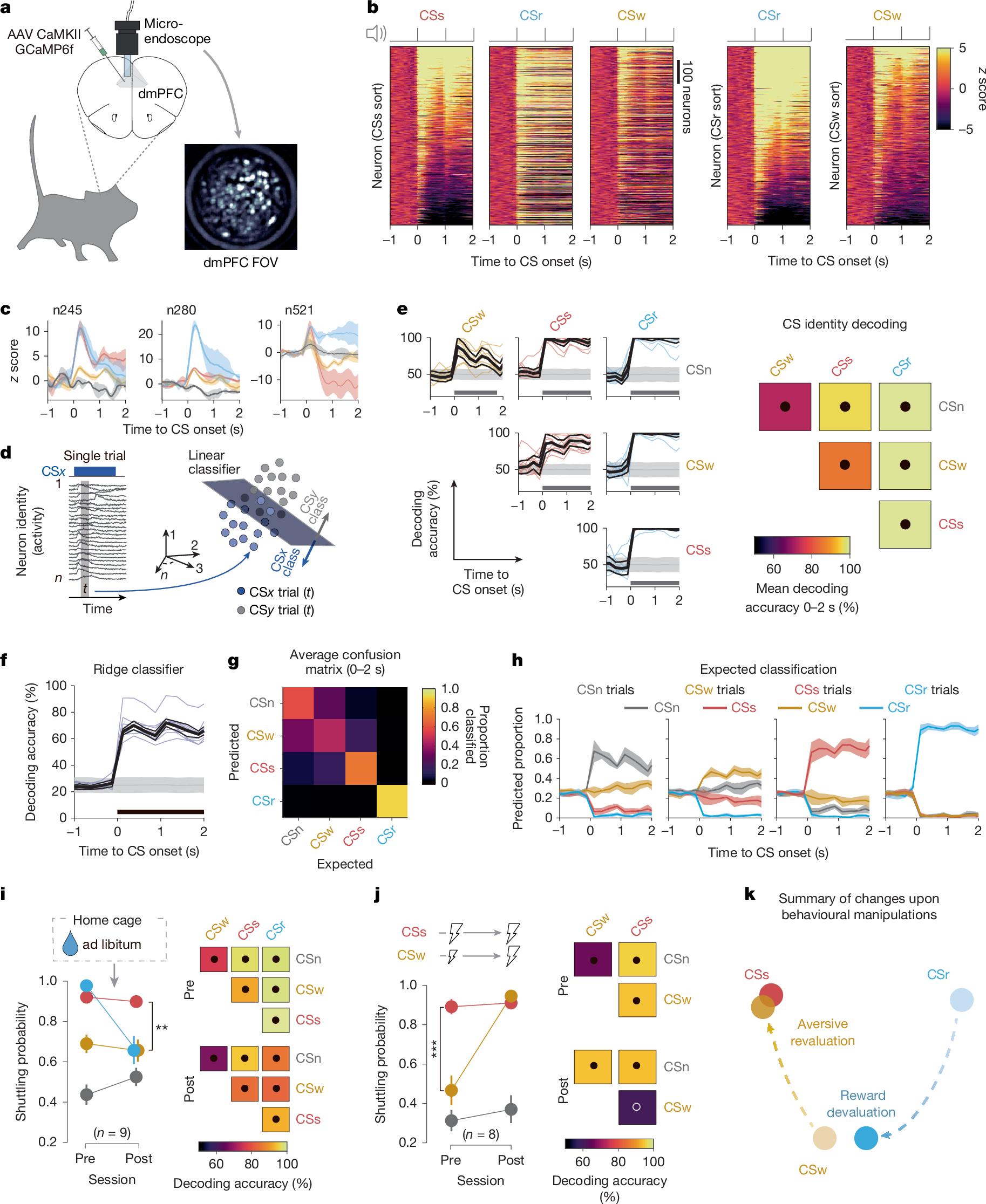
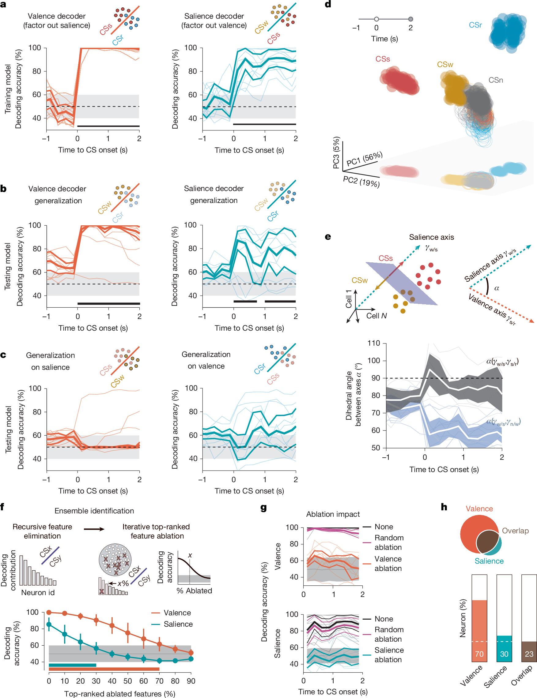
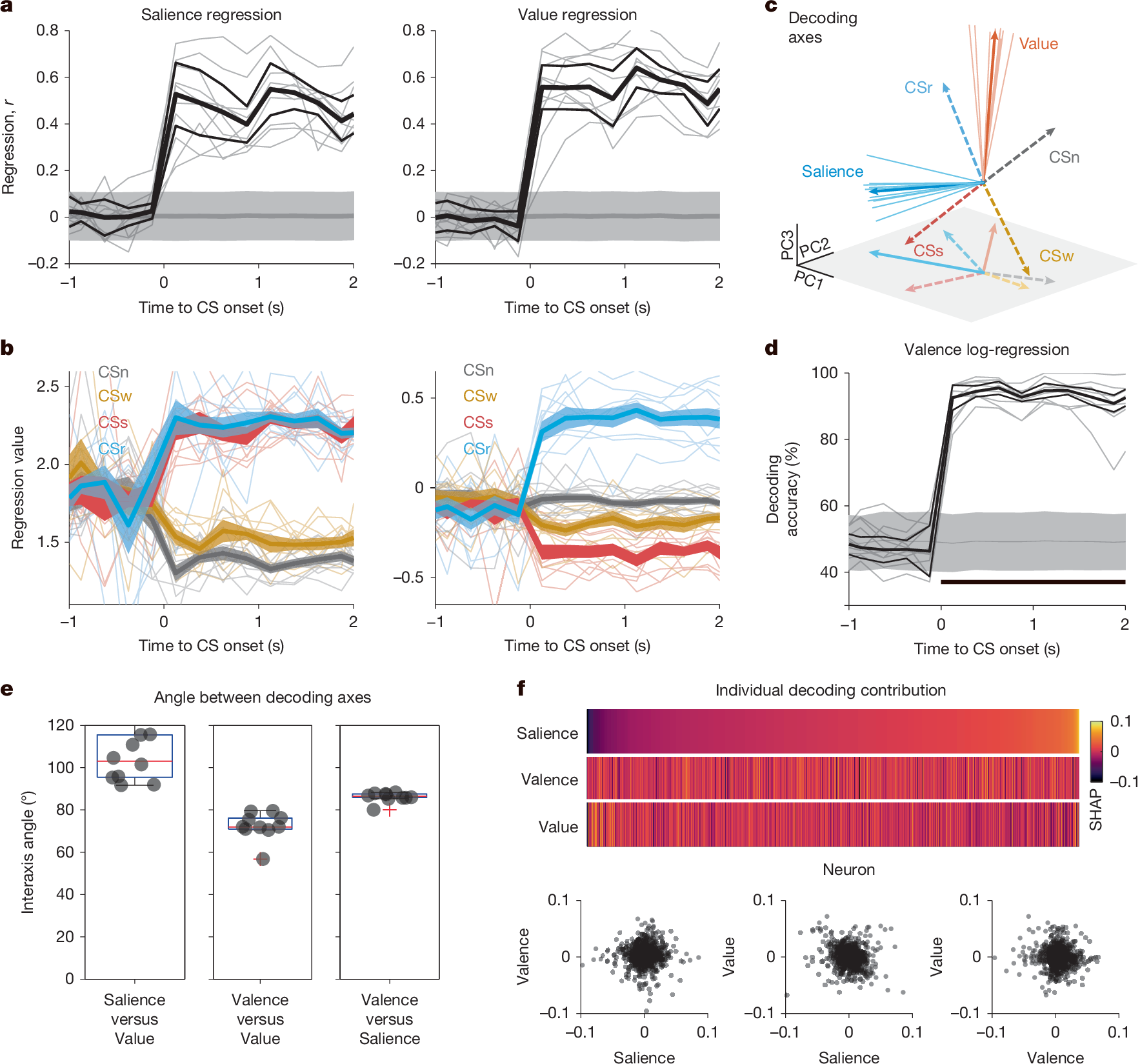
Animals continuously evaluate their surroundings to decide whether to approach rewarding opportunities or avoid potential threats. Assigning the appropriate importance to environmental stimuli is not only crucial for survival but also underlies complex forms of goal-directed behaviour that are shared across species, including humans1,2,3,4. Understanding how the brain translates such sensory cues into motivated behaviours is, therefore, central to neuroscience and psychology. The dorsomedial prefrontal cortex (dmPFC) is a critical structure that bridges relevant environmental stimuli to goal-directed behaviour. Salience, valence and value are key dimensions defining stimulus relevance, but how the dmPFC processes and organizes such dimensions to drive motivated behaviour remains unclear. Here we monitored single-neuron populations in the dmPFC using calcium imaging in freely moving male mice while discriminating between stimuli predicting different reward or punishment outcomes, which enabled an unprecedented dissociation of salience, valence and value information. We found that dmPFC populations primarily encode appetitive and aversive values of learned stimuli and that subpopulations encode valence and salience along orthogonal information axes. Our results highlight a concurrent multifaceted population coding of value, salience and valence of stimuli during associative learning within dmPFC networks, such that the geometry of dmPFC neuronal representations dynamically shapes appetitive and aversive motivated behaviours.
Access Nature and 54 other Nature Portfolio journals
Get Nature+, our best-value online-access subscription
$32.99 / 30 days
cancel any time
Subscribe to this journal
Receive 51 print issues and online access
$199.00 per year
only $3.90 per issue
Buy this article
- Purchase on SpringerLink
- Instant access to the full article PDF.
USD 39.95
Prices may be subject to local taxes which are calculated during checkout
Distributed processing for value-based choice by prelimbic circuits targeting anterior-posterior dorsal striatal subregions in male mice
Dynamical prefrontal population coding during defensive behaviours
Bi-directional regulation of cognitive control by distinct prefrontal cortical output neurons to thalamus and striatum
Data availability
Datasets that support the main findings of this study are available at GitHub (https://github.com/djercog/WinkeEtAl-value-2025).
Code availability
All data analyses were conducted using standard, built-in MATLAB (MathWorks) packages and Python libraries.
References
Pearce, J. M. & Bouton, M. E. Theories of associative learning in animals. Annu. Rev. Psychol. 52, 111–139 (2001).
Rolls, E. T. What are emotional states, and why do we have them? Emot. Rev. 5, 241–247 (2013).
O’Doherty, J. P. The problem with value. Neurosci. Biobehav. Rev. 43, 259–268 (2014).
Tye, K. M. Neural circuit motifs in valence processing. Neuron 100, 436–452 (2018).
Kahnt, T., Park, S. Q., Haynes, J. D. & Tobler, P. N. Disentangling neural representations of value and salience in the human brain. Proc. Natl Acad. Sci. USA 111, 5000–5005 (2014).
Rangel, A., Camerer, C. & Montague, P. R. A framework for studying the neurobiology of value-based decision making. Nat. Rev. Neurosci. 9, 545–556 (2008).
Rolls, E. T. Emotion Explained (Oxford Academic, 2009).
Paton, J. J., Belova, M. A., Morrison, S. E. & Salzman, C. D. The primate amygdala represents the positive and negative value of visual stimuli during learning. Nature 439, 865–870 (2006).
Article ADS PubMed PubMed Central journal=Nature&